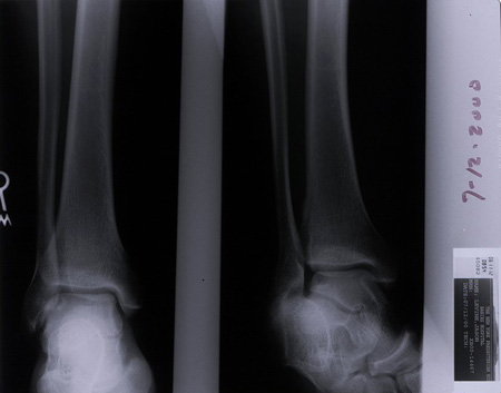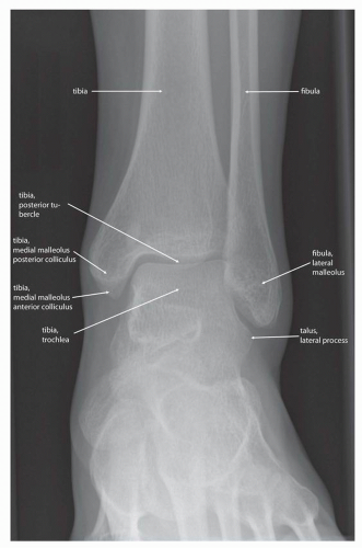

The difference in tibiofibular overlap, tibiofibular clear space and medial clear space on AP view of both sides was <50% in 97.1%, 93.1%, and 97.2% of patients, respectively. Talocrural angle, tibiofibular overlap on AP view, tibiofibular clear space on AP and mortise views, medial clear space on AP and mortise views and fibular position were significantly larger in boys than in girls. Tibiofibular overlap on AP and mortise views, relative fibular width on AP view significantly increased by age. For more information, you can read a more in-depth reference article: ankle series.


Reference article This is a summary article. It is performed to look for evidence of injury (or pathology) affecting the ankle, often after trauma.
#Normal ankle xray series#
Tibiofibular clear space on mortise views, and medial clear space on AP and mortise view significantly decreased by age. An ankle x-ray, also known as ankle series or ankle radiograph, is a set of two x-rays of the ankle joint. The timing of ossification of medial malleolus and appearance of tibial incisura between boys and girls were not different. Tibiofibular overlap, tibiofibular clear space, medial clear space, talar tilt, talocrural angle, relative fibular width and fibular position were measured.Īll radiographic measurements showed good to excellent intraobserver and interobserver reliability (ICCs, 0.603 to 0.949). If a syndesmotic ankle sprain is suspected the initial imaging choice may be 3 view radiographs taken in single leg standing. Presence of the medial malleolus and incisura fibularis were recorded. Clinical indications of a syndesmotic ankle sprain may be the mechanism of injury, pain at the distal tibiofibular joint and positive special tests (dorsiflexion external rotation test, squeeze test and cotton test). This study included 590 subjects (0-15 years), who underwent ankle AP, lateral and mortise radiographs. The standard radiographic measures presented in the present study provide the foundation for understanding the osseous foot and ankle position in a normal population.The purpose of this study was to determine the reliability of numerous radiographic measurements of the skeletally immature ankle joint, timing of ossification of medial malleolus and appearance of tibial incisura and differences in the values of radiographic measurements based on age and sex.

The radiographic angles and measurements presented in the present study demonstrate a comprehensive and useful set of standard angles, measures, and reference points that can be used in clinical and perioperative evaluation of the foot and ankle. All angles were measured by both senior authors twice, independent of each other. A total of 4 measurements were made from the axial view, 12 from the lateral view, and 17 from the anteroposterior view. The radiographic measurements were performed on standard weightbearing anteroposterior, lateral, and axial views of the right foot. A total of 33 angles and reference points were measured on 24 healthy feet. Critical preoperative planning and intraoperative and postoperative evaluation of radiographs are essential for proper deformity planning and correction of all foot and ankle cases. Objective radiographic measures are the building blocks for surgical planning. Lateral images are obtained by directing the x-ray beam from left to right, with the image receptor medial to the ankle. The limb deformity-based principles originate from a standard set of lower extremity radiographic angles and reference points. Internal oblique images are obtained by internally rotating the ankle 1520 degrees and directing the x-ray beam in a dorsoplantar direction similar to the AP view.


 0 kommentar(er)
